Our Latest Medical Blog Posts
Glaucoma is an eye disease which causes permanent and non-reversible vision loss due to rise in intra-ocular pressure (IOP) and damage to the optic nerve head at the back of the eye ball, which connect the eye ball to the brain.
It occurs very silently, and patient is normally unaware of it, till it becomes very late and vision damage is quite significant. Only a comprehensive eye examination by a qualified ophthalmologist can diagnose a case of glaucoma.
Our eye basically has two chambers, anterior chamber (in the front part) and posterior chamber (in the back part). Anterior chamber (AC) is filled with fluid called aqueous humor and posterior chamber also called vitreous cavity (VC) is filled with a jelly called vitreous humor. These fluids are essential for providing nutrition to the structure of the eye ball. Aqueous humor is continuously been secreted into AC by ciliary body epithelium and drained out via drainage angle, thereby maintaining a constant pressure inside the eye ball known as intraocular pressure (IOP). If the drainage angle is not working properly or there is some obstruction beyond the drainage angle, fluid accumulates inside AC and the IOP rises above normal.
The range of normal IOP varies between 10 to 21 mm of Hg. It is not static throughout the day and has a diurnal variation. The IOP between the two eyes is generally same or similar, within a range of ±3 mm of Hg.
Optic nerve contains more than a million tiny nerve fibers, which carries signal from the eye (retina) to the brain. Because of rise in IOP, these tiny nerve fibers die, resulting in formation of blind spots in the vision, which if not detected and treated early, will lead to total and permanent loss of vision. Because, these dead nerve fibers never regenerate, so early treatment can help in preventing further loss.
Glaucoma types:
Mainly two types: open angle glaucoma and angle closure glaucoma
Open angle glaucoma: this is the most common type of glaucoma. It occurs very slowly, and normally the affected person is not aware of it until it is very late. There is no associated pain, redness etc. in this disease. Here the aqueous humor formation is normal, but the drainage is defective, thereby causing the rise of IOP and damage to the optic nerve head.
Angle closure glaucoma: also known as narrow angle glaucoma. Here the anterior chamber is very narrow, due to iris coming forward and blocking the anterior chamber angle. Sometime due to swollen crystalline lens (cataract) iris is pushed forward from behind causing sudden and acute rise of intra ocular pressure. There is redness, severe pain, vomiting and sudden loss of vision. In some patients angle closure glaucoma develops slowly, where there are no symptoms initially. This is called chronic angle closure glaucoma. If untreated it progresses further, causing severe rise of IOP, damage optic nerve head, leading to an acute attack.
IOP measurement by Goldman applanation tonometer, OCT to screen optic nerve head to look for retinal nerve fiber layer and perimetry test to look for field of vision are important for diagnosis of glaucoma.
Comprehensive annual eye examination by qualified ophthalmologist is desirable for early diagnosis and necessary treatment of glaucoma.
Glasses / spectacles are used mostly for vision correction. However, for cosmetic purpose and protection from extreme sunlight, prevention from risk of injury etc, people regularly use eye wear. Glasses are usually called by their primary function, e.g., prescription glasses, but also appear in combinations such as prescription sunglasses, safety glasses, reading glasses, sunglasses etc. Safety glasses provide eye protection from injury in industry work, sports, post operative cases etc.
The spectacle frame is part of a pair of glasses that is designed to hold the lenses in the proper position. Ophthalmic frames come in a variety of styles, sizes, materials, shapes, and colours. Plastic, polymers, cellulose acetate, cellulose propionate, nylon, metal are different materials used for frame making. Shape of the spectacle frame are usually of round, square, cat eye, rectangle, oval, hexagonal etc. Types of frames are full frame, half frame and rimless.
Corrective lenses are used to correct refractive errors by bending the light entering into the eye. Different ocular condition such as myopia (near sightedness), hypermetropia (far sightedness) and astigmatism are corrected by corrective glasses. It brings the image back into focus on the retina. People over forty years of age suffers from difficulty in near vision, called presbyopia, which happens due to loss of crystalline lens elasticity and decreasing strength of ciliary muscle.
Types of prescription glasses are single vision glasses and bifocals. Bifocals are of different types: KB, progressive bifocal, executive bifocals, D bifocal etc. Lens material are mainly of acetate, TR90, polycarbonate and glass. Different coating present on the surface so as to make the lens anti-scratch, blue light blocking, anti-fog, anti-glare etc.
Photochromatic glasses are lightly tinted in dark in indoor conditions, but turn into deeper colours (brown, black, grey etc) when they come into contact with intense light.
An ophthalmologist or optometrist can prescribe the exact glass power required for a person, lensometer is an instrument which helps in determining the power of a person objectively.
Reading glasses offer a cheap, practical solution, though these have a pair of simple lenses of equal power, and so will not correct refraction problems like astigmatism etc.
A spectacle enhances the clarity of vision, also reduce problems like eye strain, headache etc.
Safety glasses are made with break-proof plastic lenses to protect the eye from flying debris or other matter.
Light polarization is an added feature that can be applied to sunglass lenses. Polarization filters are positioned to remove horizontally polarized rays of light, which eliminates glare from horizontal surfaces.
Sunglasses may also have corrective lenses, which requires a prescription. Clip-on sunglasses or sunglass clips can be attached to another pair of glasses. Sunglasses are used to protect one's eyes against damage from excessive levels of ultraviolet rays in sunlight. Typical sunglasses lenses are tinted for protection against bright light or polarized to remove glare. Most over-the-counter sunglasses do not have corrective power in the lenses; however, special prescription sunglasses can also be made.
Blue light overexposure are harmful to the retina, hence blue light blocking from computer, smartphone, tablets etc are increasing popular eyeglasses. It protects the eyes from dry eyes, eye strain, macular degeneration etc.
In ophthalmology (subject which studies eye in health and disease status), OCT technology is used. It is a non-invasive imaging technique which helps in visualization of the structures of the inside of the eye. It provides images of the layers of the retina, optic nerve image etc.
In diseases like diabetic retinopathy, age related macular degeneration, glaucoma etc it helps in diagnosing and following up the disease progression (worsening) while under treatment.
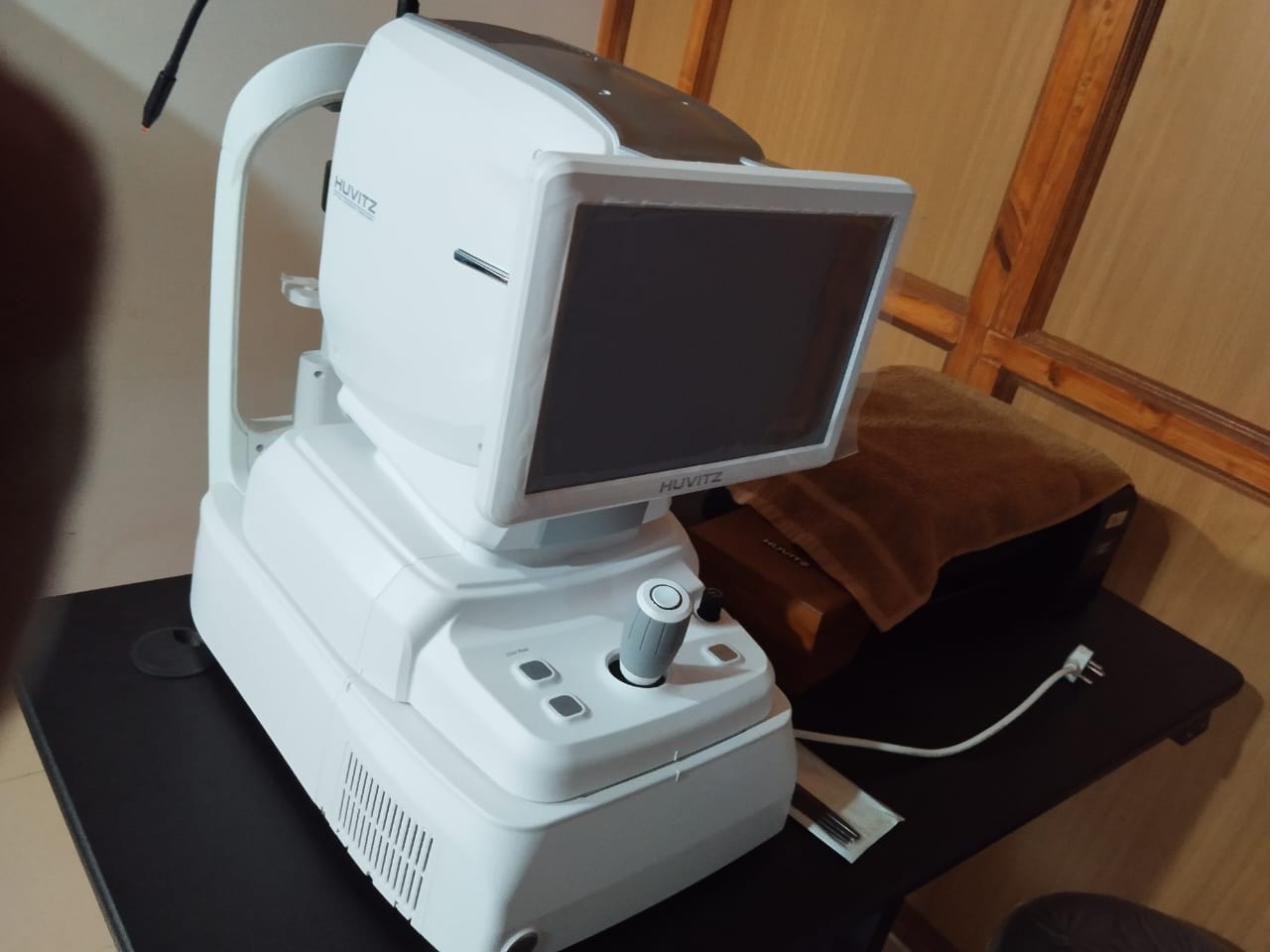
Huvitz OCT system designed specifically for ophthalmology use.
- Retinal imaging: Huvitz OCT system enable detailed visualization of the retina, including macula, optic nerve head and retinal layers. This OCT system is particularly useful for diagnosing and monitoring retinal diseases like age related macular degeration (ARMD), diabetic retinopathy (DR), macular edema (DME), retinal vein occlusion (BRVO) etc.
- Glaucoma: Huvitz OCT system can assess the optic nerve head including its cupping, thickness of nerve fibre layer. It helps in early diagnosis and meticulous follow up of glaucoma cases.
- Cornea and anterior segment scanning: It provides detail cross sectional image of the anterior segment of the eye including anterior chamber, angle, iris and crystalline lens. It also provides measurements of corneal thickness, curvature and topography. It helps in planning the placement of incision and placement of IOL in cataract surgery.
- Angio OCT: This newer modality helps in visualization of choroidal and retinal circulation without use of any dye (fluorescein etc in FFA). Helps detect and follow up of cases e.g., CNVM etc
N.B: we have the latest version of Huvitz OCT with facility of scanning macula, optic nerve, RNFL, anterior segment including cornea and angio OCT.
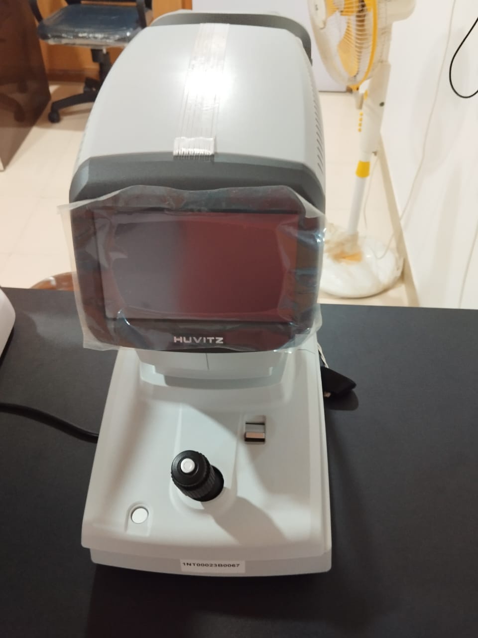
This equipment helps in measuring intra-ocular pressure (IOP) without making any direct contact with the cornea.
It is basic test done in all comprehensive eye examination. Increase IOP is called glaucoma, causes permanent damage to the optic nerve thereby causing permanent non-recoverable vision loss.
Hence NCT is a valuable instrument. Of course, applanation tonometry (AT) is the gold standard for IOP measurement,
but it is difficult to take measurement with AT in paediatric patient and very un-cooperative patient. Moreover, patients with infective diseases, IOP can be measured with NCT but not with AT.
NCT is also known as air-puff tonometer (puff of air is used). The tonometer consists of a small cone or nozzle that releases a gentle burst of air towards the cornea.
The instruments measures the force required to flatten a portion of cornea caused by the air puff and calculates the intraocular pressure (in mm of Hg) based on this measurement.
NCT is a quick and non- invasive method for measuring IOP. NCT does not requires use of local anaesthetic eye drop, as there is no direct with the cornea.
So it is convenient for routine screening in almost all types of patients for measuring IOP.
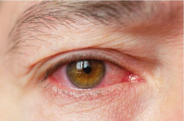
Conjunctiva is the white membranous layer that covers the eyelids and the eyeball. It is traversed by small blood vessels which are visible from outside, but with great difficulty.
If any infection affects the eye, then this conjunctiva become swollen and irritated, and it is more prominently visible from outside. This is what causes the whites of the eyes
to appear reddish or pink. This disease entity, which affects the conjunctiva is called conjunctivitis or red eye.
Red eye is most often caused by viral infection. It can also be caused by bacterial infection, allergic reaction etc. Patients with red eye complains of irritation, watering,
discharge (watery in viral infection, mucopurulent in bacterial infection)
In red eye, there is rarely any effect on vision, unless there is secondary involvement of the cornea (black circular, central structure in the front part of eyeball).
If not treated well adequately, patient suffering from red eye, will lead to loss of vision due to infection of the cornea.
Red eye is highly contagious, spread from one to another in close contact either at home or at school or work place. So, proper hygiene is very important to prevent
further spreading from one another. Towel, handkerchief etc. should not be shared between the family members.
Frequent washing of face, cleaning of eyelids with cotton tip applicator. Use of sunglasses while going outside or even at home is recommended. One should not share bed
with someone who has red eye.
Getting an early diagnosis and adequate treatment is essential. If only one eye is affected it is better to have other eye protected by cleaning and maintenance of
proper hygiene, no need to put prophylactic eye drop in the good eye, if it is not at all affected.
Urgently consult an ophthalmologist, when there is red eye, as it is a medical emergency. Any delay in doing so, may leads to serious consequences.
Directly procuring from pharmacy and applying an eye drop without consulting an ophthalmologist is highly risky and is not recommended.